Almost a century ago, a monumental breakthrough in microscopy gave the scientific community an entirely new way to peer deep inside biological cells without destroying them. Known as phase contrast, the technique transformed how researchers examined faint or nearly invisible cellular structures, making them sharply distinguishable against their backgrounds. Its profound importance to the biological and physical sciences was officially recognized with a Nobel Prize in 1953.
Now, a team of physicists at the University of California, Berkeley, has successfully engineered a way to bring that same foundational optical principle into the realm of modern electron microscopy—a technology capable of magnifying objects roughly 10,000 times more powerfully than conventional light microscopes.
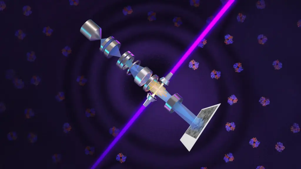
The UC Berkeley researchers have developed an extraordinary device known as a laser phase plate, an innovation that promises to significantly enhance cryoelectron microscopy, commonly referred to as cryo-EM. Over the past decade, this powerful imaging method has fundamentally transformed the study of complex protein structures and drastically accelerated the pace of pharmaceutical drug discovery. Even with its massive successes, however, cryo-EM has labored under a major, persistent limitation: the vast majority of human proteins are simply too small, meaning that obtaining crisp, clear images of them remains an extraordinarily difficult task.
The newly developed laser technology could finally help scientists overcome that stubborn obstacle. According to the research team, the system has the potential to allow scientists to image proteins as small as one-third the size of those that currently challenge modern microscopes, opening entirely new windows into molecular biology.
An even greater impact could stem from how this technology improves cryoelectron tomography, or cryo-ET. This advanced approach combines microscopic images captured from multiple angles to construct comprehensive, three-dimensional views of proteins and other vital molecules. Unlike traditional techniques that must study isolated molecules suspended in a solution, cryo-ET has the unique capacity to reveal precisely how proteins are arranged and how they physically interact within their natural cellular surroundings.
"Cryo-EM has become the new, fastest-growing method for resolving the structure of biological macromolecules, and cryo-ET is expected to show how these molecules work together in their natural, cellular context," said Holger Müller, a UC Berkeley professor of physics and faculty scientist at Lawrence Berkeley National Laboratory who spearheaded the development effort. "Pero because of signal-to-noise limitations, the majority of human and animal proteins are too small to be analyzed by these methods. The increase in signal-to-noise ratio provided by this laser phase plate is expected to overcome these important limitations."
An Extraordinary Laser Reveals Hidden Cellular Structures
The core of this scientific breakthrough relies on what the researchers describe as the world’s most intense focused continuous-wave laser. Rather than simply functioning as a light source to illuminate a specimen from the outside, the laser interacts directly and dynamically with the microscope’s electron beam, actively altering its phase in real-time to dramatically increase image contrast.
This precise adjustment makes it vastly easier to distinguish small molecules—including critical biological components like hemoglobin—from their chaotic surroundings. Furthermore, it could markedly improve the visibility of delicate internal structures inside living cells, such as the nucleus and energy-producing mitochondria.
"With cryo-ET, we’re looking at small, very complicated cellular material that’s incredibly crowded inside the cell," explained Bridget Carragher, founding technical director of imaging at Biohub in Redwood City, California. "It’s like a forest of trees, and you’re trying to find one leaf on one tree in there. Cryo-ET needs a dramatic step forward in contrast, so we can start to see what’s going on inside the cell. That’s what the laser phase plate promises to give us."
With substantial financial backing from Biohub, Müller acquired an advanced cryo-EM instrument and equipped it directly with the new laser phase plate. Fittingly, he named the resulting microscope Theia, after the ancient Greek Titaness closely associated with light, radiance, and sight.
Meanwhile, Carragher is directing the construction of a related, highly specialized microscope at Biohub’s imaging facility in Redwood City. Her team’s instrument utilizes an innovative dual-laser configuration rather than a single beam, directly building upon theoretical research previously conducted by Müller and his academic collaborators.
In this secondary setup, the two laser beams are arranged precisely at right angles to one another, with each operating at approximately half the total power. This clever configuration helps prevent delicate optical components from burning out under extreme stress while simultaneously reducing optical distortions that could otherwise interfere with the ultimate image quality.
Both independent research teams are working closely with Thermo Fisher Scientific, the undisputed global leader in the manufacturing of commercial cryo-EM instruments.
"Theia is the Formula 1 microscope," Müller remarked. "It has extra electron optics that give it better resolution than the standard cryo-EM, even without the laser. With the addition of the laser phase plate, we hope that it really becomes the world’s best instrument overall."
The Berkeley research team’s latest images and technical findings have been published in the journal Science, while comprehensive details concerning Biohub’s innovative two-laser design are detailed in a recently posted preprint.
How a Nobel Prize-Winning Discovery Changed Microscopy
To fully appreciate why this new technology matters so profoundly to modern science, it is helpful to look back at a longstanding, fundamental challenge in biological imaging.
Because plant and animal cells consist overwhelmingly of water, they are naturally transparent when viewed under an ordinary light microscope. Although this transparency might initially seem like an advantage for observing intact biological samples, it creates a severe practical problem. Small cellular structures, including nuclei and mitochondria, scatter very little light as it passes through them. Consequently, they appear only slightly darker than the surrounding aqueous material, rendering them nearly invisible.
Historically, scientists attempted to solve this visibility problem by applying chemical stains to create visual contrast. However, the harsh process of staining invariably kills the cells, making it impossible to study living cellular processes.
The paradigm shifted dramatically in 1930, when Dutch physicist Frits Zernike realized that light passing through a biological specimen changes in more ways than simply becoming brighter or dimmer. When light scatters inside a microscopic cell, it also experiences a tiny, imperceptible delay that alters its phase—the precise timing of its wave peaks.
Although the human eye cannot directly detect this subtle phase shift, Zernike discovered that it could be cleverly converted into a visible difference in brightness. By shifting the phase of the unscattered background light by exactly 90 degrees, he made subtle cellular features stand out with stark clarity against their background when the light ultimately reached the viewer’s eye.
This profound innovation became the bedrock of phase-contrast microscopy, ultimately earning Zernike the 1953 Nobel Prize in Physics.
By the early 1940s, scientists had already recognized how useful phase contrast was for studying biological cells. They quickly began exploring whether the same optical concept could be adapted to improve electron microscopes, which use focused streams of electrons instead of light waves to examine structures as unimaginably small as individual proteins.
Unfortunately, those early experimental attempts immediately encountered severe technical roadblocks. Devices designed to alter the phase of electron beams frequently weakened the beam far too much, introduced unacceptable levels of image instability, or drastically reduced the overall resolution.
A viable solution finally emerged in 2010, when Müller and Robert Glaeser—now a UC Berkeley professor emeritus of molecular and cell biology—published a proposal suggesting the use of an intense laser to produce the necessary phase shift without sacrificing electron quality.
Overcoming the Limits of Electron Microscopy
Glaeser has played a monumental role in the historical development of cryo-EM, which offers a potentially streamlined way to determine molecular structures compared to traditional X-ray crystallography.
X-ray crystallography requires researchers to grow pristine crystals of the specific molecules they want to study and gain access to an extraordinarily intense X-ray source. While cryo-EM successfully avoids the notoriously difficult requirement of crystallizing molecules, it introduces unique obstacles of its own.
One of the most destructive problems is that a high-energy electron beam can heat and eventually damage the biological specimen being examined. This radiation damage strictly limits how much data can be collected and how detailed the resulting reconstructed images can become. Although coating specimens in thin layers of metal can reduce structural damage and improve visual contrast, it unfortunately makes the final images much less sharp.
During the 1960s, researchers began investigating whether flash-freezing biological samples could protect them from radiation damage during the imaging process. Glaeser successfully demonstrated that cooling specimens to cryogenic temperatures helped preserve their native structures during imaging. He also proposed using a much less intense electron beam to examine thousands of frozen molecules simultaneously, further limiting cumulative radiation damage.
Because the individual molecules would naturally be positioned at varying angles within the ice, computers could then analyze and mathematically combine their individual projections to reconstruct a highly detailed three-dimensional molecular structure.
These pioneering advances laid the foundational framework for modern cryo-EM, the developers of which were awarded the 2017 Nobel Prize in Chemistry. During their acceptance speeches, the Nobel laureates specifically acknowledged Glaeser’s foundational contributions.
The Nobel Committee praised the technology, explaining that cryo-EM "both simplifies and improves the imaging of biomolecules. This method has moved biochemistry into a new era."
A 15-Year Quest to Harness an Exceptionally Powerful Laser
Turning the theoretical laser phase plate concept into a functional, working technology required fifteen years of grueling engineering and iterative design.
Following their initial 2010 proposal, Müller spent a decade and a half pursuing the idea, with financial backing partially provided by the National Institutes of Health. The first major hurdle was simply generating an extraordinarily intense laser beam and concentrating it into an unimaginably tiny area. The researchers needed an immense amount of light intensity to alter the phase of an electron beam by the required 90 degrees.
Their eventual solution involved confining a continuous laser beam inside a specialized spherical chamber lined with high-reflectivity mirrors. As the light repeatedly reflected back and forth through the optical cavity more than 10,000 times, its internal intensity increased exponentially while the beam narrowed into an exceptionally small focal spot.
"It’s 75 kilowatts focused to a few microns," Müller noted. "That’s more powerful than what you use for welding. It’s more power than a military laser. It builds up the brightest continuous laser focus ever."
The research team first proved the viability of their concept by installing an early prototype laser phase plate into one of Glaeser’s older electron microscopes. Subsequent funding from Biohub later enabled them to acquire a customized, state-of-the-art Thermo Fisher Krios cryo-EM instrument and heavily modify it to seamlessly accommodate the powerful laser technology.
In their most recent round of experiments, the scientists tested the heavily upgraded microscope using six distinct biological samples that varied widely in size and preparation method. The laser system consistently improved image resolution, delivering critically important gains for fragile specimens that remain notoriously difficult to image using standard techniques.
"For the most challenging cases—small particles, bad specimens—the laser produces a very considerable advantage," Müller said.
Sharper Images of Proteins Once Too Small to Study
To explicitly demonstrate the upgraded microscope’s advanced capabilities, the researchers reconstructed detailed images of two different proteins.
The first test subject was aldolase, a common muscle protein that modern cryo-EM systems can already image relatively easily. The second was hemoglobin, the vital protein responsible for carrying oxygen through the human bloodstream.
Hemoglobin is substantially smaller than aldolase and lies near the absolute lower size limit of proteins that conventional cryo-EM instruments can reliably analyze. Using the laser phase plate, the researchers successfully improved the resolution of both protein structures. The performance improvement was especially pronounced for hemoglobin, highlighting the technology’s immense potential to bypass the traditional barriers associated with smaller molecules.
"The bottom line is, if you have a large protein and a really good sample—a fresh one or one frozen without bubbles, for example—you may not need the phase plate to get a single, high-quality image. But for a small protein and a bad sample, laser-on is best," Müller explained. "This could fill an enormous gap in our knowledge of protein structures that can’t be crystallized or are too small for today’s cryo-EM. And it will be revolutionary for cryo-ET."
Protein mass is traditionally measured in daltons, a standard unit named after English chemist John Dalton, where one dalton is defined as one-twelfth of the mass of a carbon-12 atom. Conventional cryo-EM systems struggle significantly when attempting to image proteins smaller than approximately 70 kilodaltons. This limitation is particularly critical because proteins falling below this specific threshold account for roughly 90 percent of the entire human proteome—the complete collection of proteins produced within the human body.
With the introduction of the laser phase plate, researchers are now capable of imaging proteins as small as 50 kilodaltons, though pushing down to that scale remains a demanding task. These proteins are even smaller and more elusive than hemoglobin.
The Next Goal Is to Reveal Even Smaller Proteins
Looking forward, Müller believes the technology can be further refined to allow scientists to study proteins as small as 17 kilodaltons, a size comparable to myoglobin, the protein responsible for storing oxygen in muscle tissues.
Achieving that ambitious goal will likely require an additional technical enhancement: utilizing a tightly focused electron beam rather than the slightly defocused beam currently required to produce sufficient baseline contrast in the absence of the laser phase plate.
According to Müller, perfecting the laser system should eventually make it possible to obtain image contrast directly from phase changes within a sharply focused electron beam. If successful, this adjustment could double the overall contrast and signal-to-noise ratio once again, compounding the dramatic improvements already unlocked by the laser phase plate.
Such progress would make it feasible to study an exponentially larger proportion of the human body’s proteins, offering unprecedented insight into how they function and interact within the crowded, dynamic environment of living cells.
"This technology is a step function change for biology," said Stephani Otte, Biohub’s Vice President of Imaging Science. "We are going to be able to see how molecular machines operate inside the living cell, in context, for the first time. What was once invisible will become visible—and that changes everything about how we understand disease."